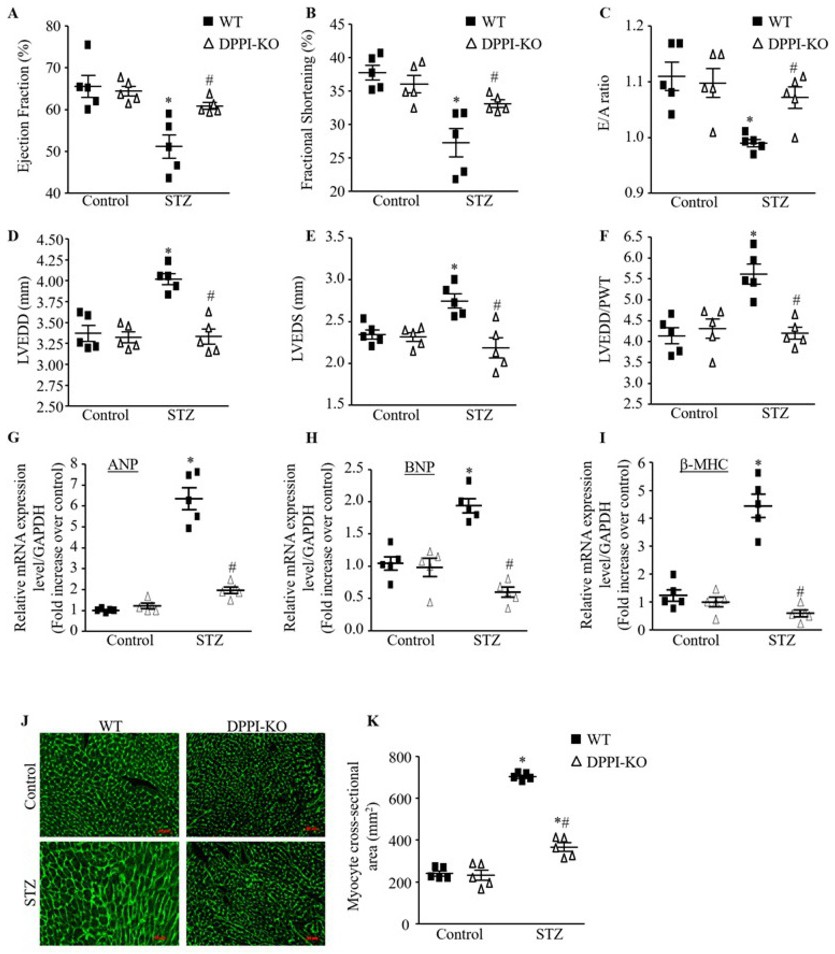
Fig. 4. DPPI deletion improve cardiac function and remodeling induced by T1DM after STZ injection independently of hyperglycemia. (A-F) Echocardiography measurement of left ventricular (LV) ejection fraction (A), fractional shortening (B), E/A ratio (C), LV end diastolic (D) and systolic (E) dimension, and LV end diastolic dimension to wall thickness ration (LVEDD/PWT) ratio (F) in control and STZ-induced diabetic animals. (G-I) Expression of cardiac remodeling markers, atrial natriuretic peptide (ANP), B-type natriuretic peptite (BNP) hypertophic markers, and beta myosin heavy chain (β-MHC). (J) Representative images of WGA staining in the controls and diabetic hearts (400X magnification with scale bars 50 µm). (H-K) Quantitative analysis of myocyte cross-sectional area. Data are expressed as means ± SEM (n=10-15; only 5 animals are shown in each group).*=p< 0.05 vs control and #=p<0.05 vs STZ-treated WT. One-way ANOVA followed by the Tukey post hoc test was used to compare multiple groups.